What Are the Common Types of MRI Scans?
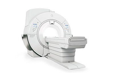
MRI scans are performed to look at the internal structures of the body. The two types of MRIs are open MRI and closed MRI. The traditional MRI involves the patient lying on a table inside a giant tube. The radio waves and giant magnets in an MRI create an image of the internal structures of the body from every angle.
Musculoskeletal MRI
Musculoskeletal MRI is a common diagnostic test used for evaluation of musculoskeletal conditions. The techniques used to create MR images have advanced over the years. Commonly used pulse sequences have also been enhanced. To properly interpret these images and to improve future imaging protocols, it is important to understand the physics involved.
Musculoskeletal MRI uses radio waves and a powerful magnetic field to produce detailed images of your body. These images are extremely sensitive and allow your doctor to accurately diagnose a variety of conditions. These images can show inflammation and bone erosion. In addition, they can help your physician determine the cause of your pain.
Musculoskeletal MRI is often part of the diagnostic algorithm for most muscle injuries. Because muscle MRI images can be easily confused with normal anatomical structures or pathological signs, interpretation of these images must be done by a multidisciplinary team. This article aims to reduce common misinterpretations of these medical images.
Before you have a Musculoskeletal MRI scan, it is important to remember that this is a procedure that requires the patient to lie still for long periods of time. Young children may require sedation to make them more comfortable during the scan. Fortunately, the process is painless and requires no blood or other invasive procedures.
Musculoskeletal MRI scans are generally performed as an outpatient procedure. The technician will position you in a narrow circular tube in order to capture images of the bones and soft tissues. This scan can be done at home or in the hospital. It can take 15 to 45 minutes to complete. The results of this test will be reviewed by a radiologist, who will interpret the results and notify your primary care physician.
Musculoskeletal radiology is a branch of radiology that deals with the imaging of bones, cartilage, connective tissue, and muscles. Its experts have special training and expertise in the diagnosis and treatment of diseases affecting the joints, muscles, and spine. They are considered leaders in their field and have a strong track record of patient outcomes.
Musculoskeletal MRI scans can be very helpful in diagnosing various disorders. Work-related disorders and sports-related injuries are a few common examples. Other disorders may include tumors in the soft tissues surrounding the joints and extremities. In addition to cancer, a Musculoskeletal MRI scan can be used to evaluate the health of the spinal cord following a trauma.
A Musculoskeletal MRI scan involves the use of a powerful magnetic field and radio frequency energy to produce detailed pictures of the internal organs and tissues. The procedure is painless and noninvasive. The results of the scan are used to plan treatment and monitor its effectiveness. The scan process usually lasts between 15 and 45 minutes, depending on the body part being examined.
The differences between a Musculoskeletal MRI scan and a CT scan are often quite similar. The differences between the two are due to the differences between water and fat protons. Therefore, understanding the differences between the two types of MRIs can help you better distinguish between a symptomatic lesion and a nonsymptomatic one.
Functional MRI
Functional MRI scans are a powerful diagnostic tool for analyzing brain functions. These scans can reveal brain structures that appear normal but are faulty, and can also reveal areas in the brain that have undergone significant changes. This type of imaging is used in hundreds of scientific articles each month, and is also often referenced in the lay press.
Functional MRI scans measure brain activity by combining multiple images taken less than a second apart. The scan works by measuring changes in blood flow within the brain during specific tasks. The results are useful for brain mapping and assessing the risk of injury and brain surgery. They can also be used to determine whether certain diseases or conditions are causing changes in the brain.
Functional MRI scans have a better spatial resolution than MEG and EEG, which are both low-resolution methods. The spatial resolution of a typical MRI is typically millimeters, but there are ultra-high-resolution scans, which can reach tens of micrometers. These MRI scans use 7-T fields and fine iron oxide to ensure the highest possible resolution.
Functional MRI scans are highly complex, requiring a lot of statistical analysis. At first, this complexity caused many to mistrust the results of functional MRI scans. However, as researchers became more familiar with the technology, these studies have become more reliable. A functional MRI scan can give a doctor a good idea of where to operate on the brain to restore function.
The MRI scanner produces a strong magnetic field, and it is recommended to avoid wearing metal items that could get in the way of the imaging process. Moreover, the area of the body that will be imaged will be slightly warm. If you experience any discomfort, it is best to inform the radiologist as soon as possible. For the best results, the patient should be perfectly still while the images are being recorded. The entire procedure typically takes a few minutes.
A functional MRI scan is an imaging test that measures brain activity by using blood oxygen level-dependent contrast. During the test, the blood in the brain is oxygenated and deoxygenated, which creates differences in the magnetic susceptibility of these two types of blood.
Functional MRI scans are commonly performed as a part of brain activity research. In the most common fMRI scans, different slices of the brain are acquired at different times. This allows researchers to compare the signals recorded during different states. The results of the analysis are used to create an activation map of the brain.
In contrast to fMRI scans, functional MRI scans are noninvasive and do not involve any surgery or injections. Rather, they are a valuable part of brain mapping research.
Open MRI
Open MRI scans are similar to closed MRIs but are more comfortable for patients. Patients who are claustrophobic or who are sensitive to loud noises may find them uncomfortable. However, newer machines are getting closer to producing images of the same quality as closed MRIs. Patients still need to lie still, and the open MRI scan type does not completely enclose the patient.
Open MRI machines are more accommodating to larger patients with claustrophobia. These MRI machines have front and back windows and are usually open on two sides. They are also more advanced than closed MRI machines, but take longer to produce images. Patients should consider the size and comfort of the MRI machine before choosing it.
Open MRIs are not as useful for detecting small or delicate body parts. But they are useful for detecting lesions in the brain and wrist, and can provide doctors with a more detailed diagnosis. They can also be used to detect single or multiple vessels in patients with coronary artery disease.
While an MRI scanner does not use radiation, the magnetic field is intense. The electromagnetic waves that travel through the body cause the atoms to temporarily move. The atoms emit different amounts of energy, which vary depending on the type of tissue. The MRI scanner captures the energy generated by the hydrogen atoms and converts it into a picture. The images can help diagnose any problems in a person's body.
Open MRI scans are quieter than closed MRIs and are also better for larger patients. Open MRIs are also more child-friendly, and a parent can be present when a child is having an MRI. They are also safer than closed MRIs.
Open MRI scans are commonly used for a variety of purposes. These types of MRIs are very useful for identifying symptoms of stroke, heart diseases, disc disease, bone infections, and more. They are also useful for diagnosing internal organs. And unlike traditional MRIs, they are able to produce more detailed images in less time.
Open MRI scans can be performed on people with claustrophobia, and the procedure is more comfortable for the patient. As the MRI machine is a circle, the patient can move into the circle's center opening, making it 20% larger than conventional MRI scanners. Additionally, most patients are able to lie on their feet while receiving the exam, making it more comfortable for them.
MRI scans highlight contrasts in soft tissues, and this makes it easier for physicians to identify problems in joints, cartilage, tendons, and ligaments. They also help identify infections and inflammatory problems. They can also help physicians rule out tumors. These scans are also useful for assessing the anatomy of the brain.
Open MRI scans can be performed in a variety of positions, and are the most common MRI scan types. There are a number of pulse sequences available for these types of imaging.


