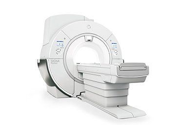
The Benefits Of A Positron Emission Tomography Scan
During a PET scan, the body's cells emit radiation called positrons, which are radioactive elements that can be used to detect disease. This method is noninvasive, painless, and enables physicians to detect disease at an early stage. It also helps detect areas of activity inside the body.
Noninvasive
A Noninvasive Positron Emission Tomography (PET) scan uses a small amount of radioactive tracer to visualize the blood flow and function of heart muscle tissue. The tracer is injected into a vein in the arm and travels to the heart where it is detected by special cameras. The process produces images of the heart at rest and when the patient is given medication. It helps doctors diagnose certain heart conditions and certain types of cancer.
A PET scan is noninvasive and generally painless. The procedure involves a small dose of radioactive material that is attached to a common compound. This is usually glucose, though other compounds similar to glucose can also be used. The patient receives the tracer through a vein and a specially designed positron scanner uses the images to obtain detailed information about the body's metabolism of the tracer. PET has been in clinical use since the early 1990s.
The most common tracers used in a PET scan are rubidium-82 (Rb) and nitrogen-13-ammonia (N-ammonia). This procedure takes less time than a SPECT scan, and the radiotracers used in a PET scan have shorter half-lives than the SPECT scan. The test can be completed in less than 30 minutes.
The main goal of a PET scan is to diagnose the cellular changes that occur in the body during disease. The PET scan uses a small amount of radioactive material to create images. The radioactive tracer is similar to sugar but is attached to a small radioactive atom. The radioactive substance travels through the bloodstream to the patient's brain and is detected by brain cells as positrons.
A PET scan is an excellent diagnostic tool. It can show whether a patient is suffering from cancer and whether it has spread throughout the body. It is also an important part of a patient's care, and can help doctors plan treatment options early.
Painless
A Painless Positron Emission Tomography (PET) scan is a type of medical test in which a radioactive substance is injected into the body through a small plastic tube. The patient lies still on the scan table for 30 to 60 minutes. The radioactive substance, known as FDG, travels through the blood to the area of the body being studied. The substance is then absorbed by the tissues, and the scan can take place.
This scan provides doctors with a detailed view of the human body and organs. It helps determine organ function by measuring the metabolism of organs and tissues. It also allows physicians to see how cancerous cells affect various organs. It is also an effective tool for detecting brain tumors and heart disease.
A PET scan uses radioactive substances to create images of the body's organs and systems. It is most commonly used to detect cancer, monitor the progression of the disease, and determine the effectiveness of chemotherapy. But it can also be used to diagnose a variety of other diseases, including cardiovascular diseases and brain disorders.
PET scans are relatively painless. They involve injecting a tracer liquid into a vein, usually in the arm. The patient may also swallow a tracer or breathe it in. The liquid then travels throughout the body and collects in an organ. As it moves through the body, it gives off positive-charged particles (positrons). A camera records the positrons, turning them into pictures on a computer screen. PET tests can help doctors assess the effects of trauma on the brain, assess the level of perfusion in the heart muscle, and assess the spread of heart disease.
Unlike other types of tests, a PET scan does not require anaesthesia or sedation. Patients need to be completely still for the procedure to be performed. PET scans are generally outpatient procedures. This means that a patient doesn't have to stay overnight in the hospital. However, patients are encouraged to follow up with their doctor if they are ill before their appointment.
Can detect disease in its earliest stages
Using a special camera and a substance called a "tracer," a positron emission tomography scan can detect cellular changes in the body. This procedure is used to diagnose heart, lung, and brain conditions. Compared to other imaging tests, the images from a PET scan can detect disease in its earliest stages.
The process of positron emission tomography involves injecting a small radioactive material into the patient's body. This radioactive substance helps the doctor create a 3-D picture of the organs in a person's body. These scans can detect the earliest stages of many types of disease and help doctors determine the effectiveness of treatment. The scans can also reveal abnormalities in organs and parts of the body, including tumors and brain disorders.
The technology behind positron emission tomography is based on the physical properties of isotopes. They emit positrons during decay. A PET center uses special radiochemical laboratories to produce these isotopes. These radioactive substances travel through the bloodstream and enter the patient's brain. Once inside, they bind with ligands in brain cells and then release positrons into the body.
In a PET scan, a positron emitter is passed through a detector pair. These detectors detect coincidence events in the positrons emitted. They are made from bismuth germinate or lutetium oxide and contain photomultiplier tubes. Signals from these detectors are then fed into separate amplifiers and energy discriminating circuits. This technique is able to pinpoint disease in its earliest stages.
In addition to the earliest detection of disease, PET scans can also help in the treatment of a variety of diseases, including cancer. PET scanners can detect disease at its early stages by measuring the metabolism of cancer cells. The FDG-F-18 positron emitter is an important workhorse in tumor imaging. Tumor cells increase their expression of glucose transporter molecules and hexokinase in their tissues, which makes it possible for them to consume more FDG.
Can detect radioactive glucose
A Positron emission tomography (PET) scan can detect cancer by detecting the levels of radioactive glucose in the body. It works by using a small amount of radioactive glucose, known as a tracer, to produce computerized images of the body's chemical structures. PET scans can detect cancer in several ways, including tumor detection, and they can be used in conjunction with a CT scan to enhance the accuracy of detecting abnormalities.
A PET scan uses a radioactive substance called FDG, or fluorodeoxyglucose, to detect tumours and other medical conditions. The substance accumulates throughout the body and gives off gamma rays that are detected by the PET scanner. Once the scan is complete, patients slide out of the machine. They can normally return to their daily activities, though they should limit their activity for 30 to 90 minutes after the scan to reduce the risk of radiation exposure.
Patients should avoid eating and drinking for several hours before the scan to avoid affecting their sugar metabolism. Patients should also refrain from strenuous exercise 24 hours before the appointment. The appointment letter will include instructions on how to prepare. The radioactive tracer is only active for a brief time, so patients should avoid eating or exercising for at least 6 hours before the scan. Patients should wear loose-fitting clothing and take off any metal jewelry before the scan.
The PET scan takes 30 to 60 minutes. After the patient is ready, he or she will be given a contrast infusion through an IV or CVC. This medication is then slowly infused into the patient's bloodstream. The patient must remain silent and still for 30 to 60 minutes before the scan. Afterwards, the doctor will look over the results. The patient can be told within a week of the scan.


