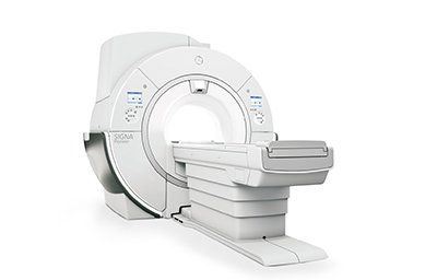
The Benefits of an MRI
An MRI scan is a safe, painless test that can show the inside of the body. It helps doctors distinguish different types of tissue because protons in the tissue realign differently and produce different signals. The signals from millions of protons combine to create a detailed picture of the interior of the body. Patients should not feel any discomfort during the test and can even lie down during the procedure. However, those who suffer from claustrophobia may need to ask a radiographer how to deal with their fear of the process.
MRI scans are non-invasive
An MRI scan is a type of imaging test that uses magnetic resonance imaging (MRI). An MRI scan doesn't involve radiation or surgery, and is therefore a great option for many patients. However, this type of test is not without risks. It can take up to an hour to perform, and the patient must remain still during the entire procedure.
An MRI scan can reveal problems that other tests cannot detect. It can be a valuable tool for the early diagnosis and evaluation of many focal lesions. The technology is extremely sensitive and can detect abnormalities that are hidden by bone. In addition, MRI scans are excellent for assessing the biliary system, as they can reveal abnormalities in this area that aren't visible on other types of imaging.
Although an MRI scan is very safe, it is important to note that there are some potential risks. The FDA receives about 300 reports of adverse events per year, and most of these describe problems related to heating or thermal injury. The most common problem reported by patients is second-degree burns, but other issues include projectile events and falls. It is also important to note that some patients experience nerve stimulation during the MRI.
MRI scans are not dangerous, but they can be stressful for people who have an extreme fear of enclosed spaces. However, sedation and other medications can be used to keep patients calm during the scan. Patients should follow the instructions of the facility to minimize potential side effects.
MRI scans are also useful for tracking treatment. They can reveal changes in organ, lesion or tumor size. In addition, they can reveal abnormal blood flow in organs. This information can help doctors diagnose suspected tumors and determine how best to treat them. An MRI scan can reveal differences in water content and blood flow between different tissues. A cancerous tumor may have an abundance of new blood vessels that supply the tumor with more blood than the surrounding tissue.
They are painless
An MRI is a safe and painless procedure. Patients are usually given a gown and asked to remove any jewelry or glasses. The exam may take a few minutes to complete. Before the test, patients may be given a sedative or anesthesia that will help them relax. A nurse will be present to help them. After the exam, patients will be taken to the recovery room.
While an MRI is a painless procedure, you will need to lie still during the procedure. If you move during the scan, the images will be blurred. The scan itself will last about 15 minutes. Some patients may experience claustrophobia, so earplugs or headphones can be helpful.
Patients should inform their physician if they are pregnant or have any metal implants before getting an MRI. Although an MRI does not cause any pain, the procedure is noisy, so it is important to wear headphones or earplugs. Patients should also remove all magnetic objects from their body before going into the machine.
The procedure itself will last between half an hour. After the procedure, the patient can resume normal activities. However, people who are afraid of being in a closed space can ask their healthcare provider about medication that will help them relax. A mild sedative will help you sleep, but it is necessary to follow all instructions closely.
During the procedure, patients will lie on a table that slides inside an MRI machine. The table may be equipped with a plastic coil that wraps around the head. The technician will then take several pictures of the brain. Each picture takes a few minutes. The patient will be able to communicate with the technician through a microphone.
They are safe for people who are vulnerable to radiation
While there are some concerns that MRIs are not safe for people who are vulnerable to radiation, some studies have shown that they are safe. This study involved a nationwide survey in England where 85 patients with leukemia and 45 unaffected controls were compared. The results showed that the difference between the two groups could not have been fortuitous. However, the study also indicated that radiation from MRIs may initiate changes in a fetus or young child.
The risk of cancer from imaging-based radiation is small compared to the overall risk of cancer. It is estimated that the risk of cancer from a single CT examination is approximately 42 per 100 patients. These small risks must be multiplied by a large population to find the overall risk. Even if the patient's dose is not excessively high, the risk of radiation-induced death is not high, averaging about 50 per 100,000 CT patients.
In 2003, the FDA classified MRIs up to 8 T as nonsignificant risk devices for nonneonatal patients. During the same period, the International Commission on Non-Ionizing Radiation Protection determined that acute exposures at this level caused no serious health effects. In addition, the International Electrotechnical Commission increased the static magnetic field limit of the first-level controlled operating mode to eight T. One vendor even received a CE mark for a 7-T clinical system.
People who are sensitive to radiation should be aware that MRIs are safe for those with genetic conditions. Because MRIs are painless, claustrophobia or other medical conditions shouldn't interfere with the scan.
They can show both bones and soft tissues
MRI, or magnetic resonance imaging, is a diagnostic test that creates cross-sectional images of your body. These images are created by a computer and magnetic field. They are a good way for doctors to view both soft and hard tissues in the body. MRIs are more detailed than x-rays and can show diseased tissues and tumors.
The radiation used during x-rays is absorbed by dense matter, such as bone, but passes through less dense soft tissues. In a typical x-ray, several angles are taken and the images are compared to those of the uninjured limb. The entire process usually takes about 10 minutes. The images are then developed and written to a CD, or can be viewed on a computer screen.
MRIs are also useful in diagnosing bone injuries. They provide detailed images of the bones and soft tissues in the body, such as the cartilage. The scan also helps doctors identify bone spurs, which are often difficult to detect with X-rays alone. An MRI can also show inflammation of soft tissues, which are not possible to detect through X-ray images.
Before an MRI, patients must be completely still and remove any jewelry or credit cards. Patients should also bring any relevant X-rays and an insurance identification card. An MRI can be difficult for people who are claustrophobic, but an open MRI can help you relax.
MRIs use contrast media to improve the visibility of soft tissues and improve diagnosis. The contrast agent is not the same as the dye you might use at home. MRIs use non-iodine gadolinium-based contrast agents. They're safe and effective. But some people may be sensitive to these drugs and a kidney function test may be required. In addition, metallic substances in the body can affect MRI images and cause discomfort or injury. Therefore, it's a good idea to consult with your physician about any medical history before scheduling an MRI.
They are faster than X-rays
An MRI is a diagnostic imaging procedure that uses radio waves and a magnetic field to create detailed pictures of internal organs, bones, and tissues. While X-rays are more commonly used to detect broken bones, an MRI can also detect diseased tissues, such as tumors. Furthermore, the radio waves used in MRIs are far safer than x-rays because they carry much lower levels of energy than visible light rays.
Another major difference between an X-ray and an MRI is the speed of scanning. An MRI can take only a few seconds, while an X-ray can take minutes. And because MRIs are faster, they are also more sensitive. This means that they're often more useful in diagnosing cancer, especially in young children. Moreover, MRIs are safer and cheaper for adults, too.
The MRI is also faster than a CT scan. CTs use ionizing radiation to create a 3D image of a region, and they are also less expensive than X-rays. X-rays are better for certain types of conditions, such as fractures and serious injuries, while MRIs are better for diagnosing internal organs, such as kidney disease.
X-rays are faster than MRIs, but they're not without their own disadvantages. Patients must lie motionless for an MRI to give the best images. This is because movement may cause blurry images. Depending on the type of scan, MRIs can take as little as 15 minutes or as long as an hour.
The speed of MRIs has increased dramatically in the last decade. The fastest brain scans can now take less than a half second. The newest MRI technology combines x-rays with a computer. This allows researchers to get a three-dimensional picture of the brain in less than a half second, compared to two to three seconds for the typical X-ray. This speed is critical for neuroscience.


