Types of Medical Imaging
Medical imaging uses a variety of techniques. These include X-rays, Computed tomography, MRI, sonography, and more. You can find out more about these types of tests below. These types of tests help doctors diagnose certain conditions by producing detailed images that a standard X-ray cannot.
X-rays
X-rays are used in medical imaging for a variety of purposes. The radiation produced by an X-ray machine passes through most solid objects in the body to produce an image. This image is then used to help healthcare providers visualize internal structures of the body. Some radiographs include contrast medium, which is given to patients intravenously, orally, or rectally. These contrast mediums are usually colored white or grey, depending on their density. Bone and other solid objects appear white, while skin, muscle, blood, and fat are gray.
Although X-rays are a common imaging modality, the risk of exposure to ionizing radiation remains low. There are a number of laws in place to ensure the safe use of X-rays. These laws are implemented by various government regulatory agencies and advisory bodies. Understanding these laws may help radiologic technologists make sound decisions regarding the use of x-rays in medical imaging.
Although X-rays are safe for most people, they may increase the risk of developing cancer if repeated exposures are made over the course of a lifetime. Therefore, it is important to discuss your risk with your healthcare provider before scheduling an appointment for an x-ray. Additionally, it is important to remember that pregnant women should never hold their babies during an x-ray.
A number of x-ray imaging methods are now available. Single-frame x-ray tomosynthesis (SRS) captures up to 30 images per second and provides a 10 to 100 times higher spatial resolution than conventional tomosynthesis. This method could lead to a safer, less invasive way to treat lung tumors and cardiovascular diseases.
X-rays are electromagnetic radiation with a very short wavelength and high energy. As such, they can penetrate through bones and soft tissue. In addition, they don't carry a charge. Unlike visible light, x-rays are not affected by magnetic fields, so they do not interfere with magnetic field measurements. They are also capable of producing a photoelectric effect, which makes them a popular tool for medical imaging.
Today, medical imaging is the largest application for X-rays. It is used for image-guided therapy and medical diagnostics. The technology behind X-rays is based on vacuum electronics and a mechanism known as bremsstrahlung. It was first discovered more than 120 years ago, and since then, it has become a valuable medical imaging tool.
MRI
MRI medical imaging is a common medical procedure, but there are certain precautions patients should take before having the procedure. For instance, pregnant women should let their MRI technologist know if they are pregnant, and should leave jewelry and metal at home. They should also bring a photo ID and insurance card.
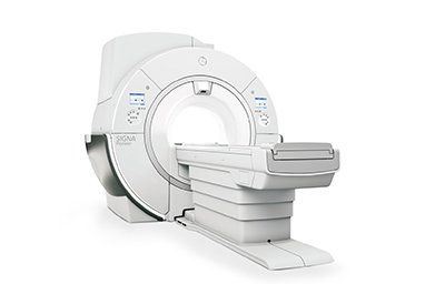
The MRI machine is a tunnel-like device that has a powerful electromagnet. This magnet forces the hydrogen atoms in the patient's body to align with its magnetic field. High-frequency radio waves are then emitted from the magnet, which causes protons to spin, and then attempt to realign with the magnetic field. This motion causes an electric signal, which is captured by a sensor.
If you're pregnant, it's best to let your MRI technologist know before you have your exam, because some of the contrast material is toxic to unborn children. It's important to disclose this to the technologist so that he can advise you on how to safely receive the contrast agent.
MRI medical imaging is often used to diagnose and treat a variety of conditions. Different MRI sequences may be useful for different purposes, such as assessing the cerebral cortex, identifying fatty tissue, and characterizing focal liver lesions. Some MRI sequences can be performed simultaneously using these techniques.
MRIs can be used to diagnose conditions such as herniated spinal discs, multiple sclerosis, and liver cirrhosis. These images are acquired in a special digital format called DICOM. DICOM ensures high-quality images. In addition to the quality of the images, MRIs can provide highly accurate data.
Before undergoing an MRI medical imaging procedure, patients must remove all metal objects from their bodies. They should also wear a cotton gown. During the procedure, patients are required to remain still. A radiography technologist monitors the process from a separate room. Patients can talk to the technologist via a microphone, which helps ease the fear of being confined in a narrow tube. The procedure can take anywhere from 15 minutes to an hour.
MRI medical imaging is typically painless. The duration of the exam varies depending on the type of study that a patient needs. The actual test time is between 20 and 60 minutes, though prep time can extend the appointment time. The time may also be increased if multiple studies are needed.
Sonography
A sonogram is a form of medical imaging that uses ultrasound waves to visualize internal structures. The sonographer uses a transducer that moves over the patient's skin and then captures images. The images are then displayed on a computer screen. The sonographer will review the images with the radiologist, who will interpret the results and provide the doctor with a report. The patient will be asked to remove any clothing that may be obscuring the area being examined. Then, the sonographer will ask the patient to lie on a bed.
There are many types of sonography, including vascular and neurosonology. Vascular sonographers create images of blood vessels and arteries, while obstetric and gynecologic sonographers focus on the female reproductive system. Other specialized sonographers specialize in imaging the abdominal and musculoskeletal systems, as well as the heart. Cardiovascular sonography focuses on cardiac arteries and valves.
The benefits of sonography are many. For instance, it can help doctors diagnose pelvic masses, infertility problems, and inflammatory diseases. It is considered one of the most reliable imaging methods. It helps doctors make accurate diagnoses without the use of radiation, surgery, or dyes. Plus, sonography is safe, portable, and non-invasive.
Sonography uses sound waves to visualize soft tissue and bone surfaces. It can also be used to diagnose muscle strains and ligament sprains. It can also be used in conjunction with x-ray imaging to detect fractures. Moreover, it can be used to guide a needle during injections.
A sonographer must be trained in the use of sophisticated computer equipment. They must also be skilled in working with patients. They must be knowledgeable about anatomy, ethics, and how to communicate with patients. They also have to develop rapport with patients and display confidence throughout the procedure. During the imaging process, a sonographer must have a strong sense of confidence and competence.
Another benefit of sonography is the real-time images it produces. This technology is much safer than X-rays, because it does not expose the patient to ionizing radiation.


